Photoacoustic imaging keeps proving itself and keeps waiting for the clinic
Light in, sound out: optical contrast at ultrasound depth. The physics is settled and the clinical adoption path is the hard part.

Photoacoustic imaging illuminates tissue with a short laser pulse. Absorbing structures heat, expand and emit an ultrasound wave, which conventional ultrasound transducers detect. The image carries optical absorption contrast at depths where purely optical imaging fails because of scattering.
Why the combination is attractive
Optical methods offer excellent contrast — haemoglobin, melanin, lipids and water all absorb distinctively, and oxygenated and deoxygenated haemoglobin differ enough to map blood oxygenation. But light scatters, so purely optical imaging loses resolution within about a millimetre of tissue.
Ultrasound penetrates centimetres with good resolution but has poor soft-tissue contrast.
Photoacoustic imaging uses light for contrast and sound for spatial information, which sidesteps the limitation of each.
What it can show
Vascular structure without contrast agents. Blood oxygenation, from measurements at multiple wavelengths, which is a functional parameter few modalities provide non-invasively. Tumour angiogenesis. Inflammation. In principle, these support oncology, dermatology, rheumatology and vascular assessment.
Why clinical adoption is slow
Depth versus resolution. Deeper imaging requires lower ultrasound frequencies and therefore coarser resolution, and light delivery becomes the limit. Practical depth is a few centimetres, which suits some anatomy and excludes others.
Quantification. Converting signal amplitude to absorber concentration requires knowing the light fluence at depth, and fluence depends on the tissue's own optical properties — which are what is being measured. Quantitative photoacoustics remains an active research problem.
Laser requirements. Wavelength-tunable pulsed sources with sufficient energy have historically been bulky and expensive. Diode and fibre-based sources are improving this.
Clinical evidence. Adoption requires trials demonstrating that the information changes patient outcomes, not merely that it can be acquired. That is slow and expensive, and it is where most novel imaging modalities stall.
Where it stands
Systems have clearance for specific indications in several jurisdictions, particularly in breast imaging and dermatology, and handheld probes integrating with clinical ultrasound have appeared.
The pattern is characteristic of medical imaging: the physics was demonstrated decades ago, and the translation timeline is governed by evidence generation and reimbursement rather than by technical capability.
This article was produced by the LasersNews AI desk and reviewed by our editors.
Related reading
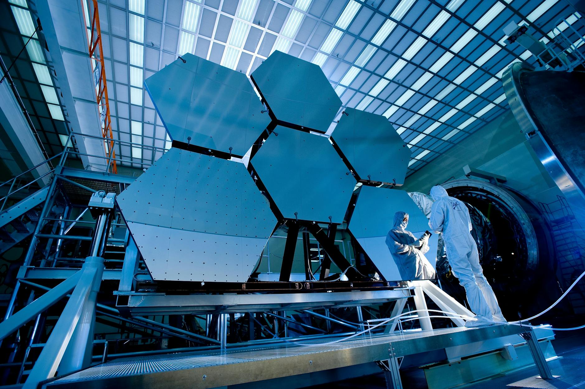
Integrated photonics narrows the gap between laboratory result and manufacturable part
Foundry access and standardised process design kits are doing for photonics roughly what they did for electro…
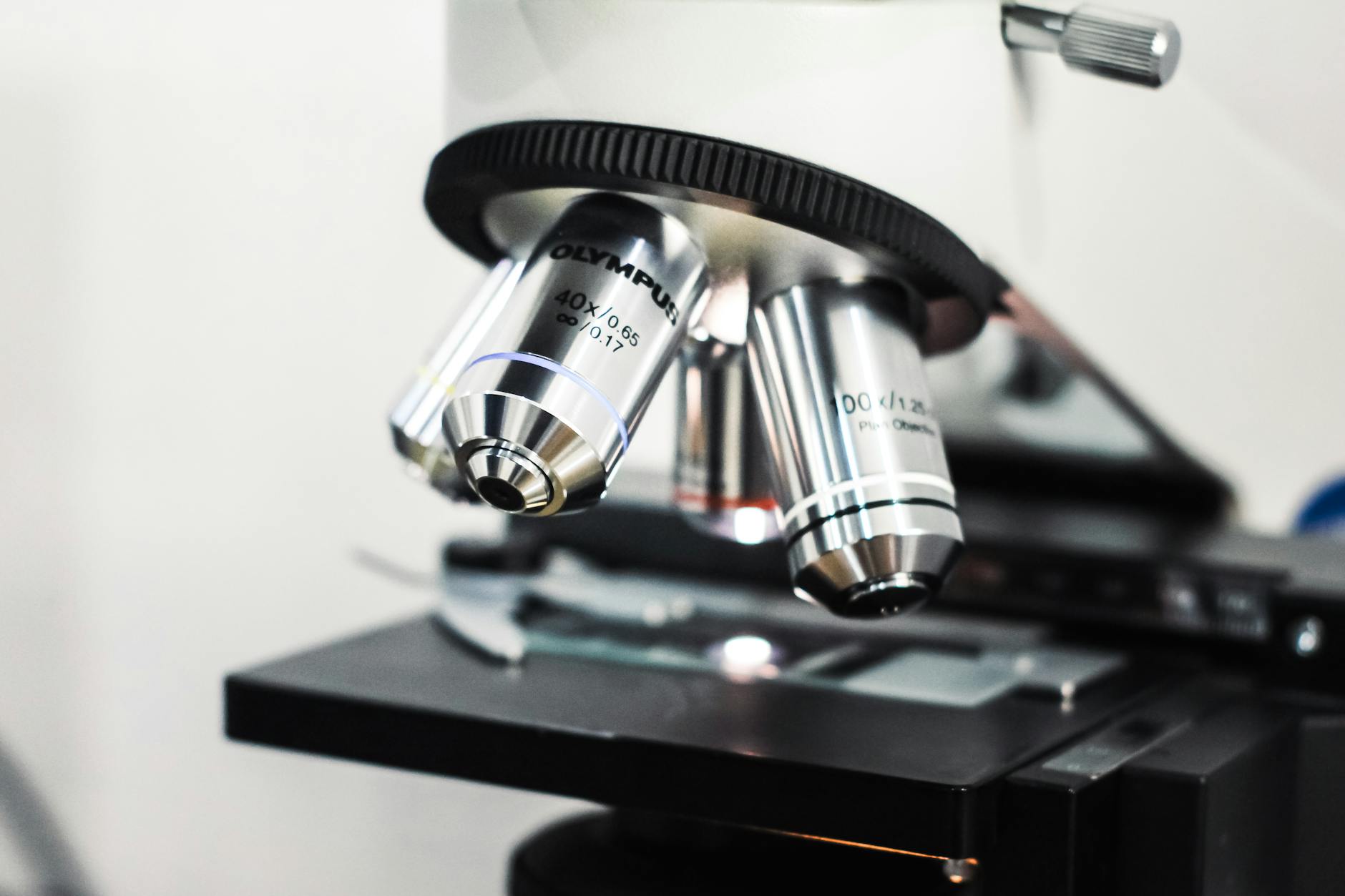
Metasurface optics are running into the manufacturing question
Flat lenses built from sub-wavelength structures work. Producing them at volume, at yield, with the tolerance…
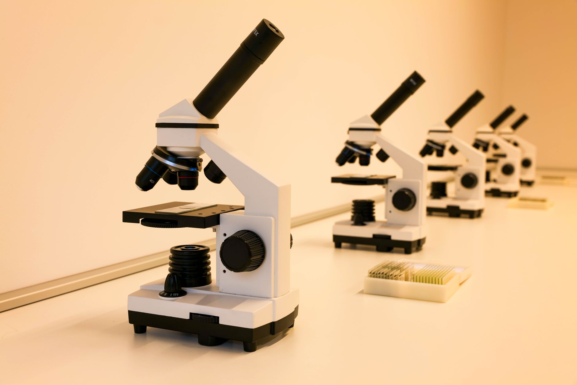
Photonics found its foundry model, and the effect resembles electronics in the 1980s
Shared process design kits and multi-project wafer runs let a small team design a photonic chip without ownin…